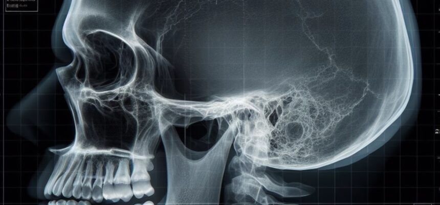
Radiographic Evaluation of the Cranium
Understanding the essentials of cranial X-ray interpretation is crucial for medical students. Here’s a concise guide:
Indications:
- Head Trauma: Assess for fractures, skull base injuries, or foreign bodies.
- Suspected Intracranial Pathology (when advanced imaging is not available): Evaluate for tumors, hydrocephalus, abscesses, or vascular abnormalities.
- Chronic Conditions: Monitor conditions like hydrocephalus or skull abnormalities.
- Follow-Up: Track progress post-surgery or intervention.
Contraindications:
- Pregnancy: Minimize exposure, especially during the first trimester.
- Unnecessary Exposure: Avoid when clinical history and physical examination provide sufficient information.
- Advanced Imaging Needed: If CT or MRI is indicated for detailed assessment.
Interpretation:
- Fractures: Look for linear fractures, depressions, or signs of healing.
- Soft Tissue: Assess for swelling, foreign bodies, or gas in the scalp.
- Skull Abnormalities: Identify congenital anomalies or bony growths.
- Sutures and Fontanelles: Evaluate the closure status in infants.
- Foreign Bodies: Check for presence and location.
Potential Findings:
- Fractures: Linear, depressed, or basilar fractures.
- Skull Abnormalities: Enlarged sella turcica, thinning of bone.
- Tumors: Presence of abnormal masses or calcifications.
- Hydrocephalus: Enlarged ventricles.
- Foreign Bodies: Presence of metallic objects or projectiles.
Actions to Be Taken:
- Consult with Radiologist: Seek expert opinion for complex cases.
- Clinical Correlation: Correlate findings with clinical history and physical examination.
- Further Imaging: If X-ray is inconclusive, consider CT or MRI for detailed assessment.
- Timely Reporting: Communicate findings promptly for appropriate patient management.
- Consider Patient Factors: Age, comorbidities, and clinical presentation influence the approach.
Verified by Dr. Petya Stefanova
