
What are pathological reflexes?
Pathological or primitive reflexes are commonly observed in infants up to approximately 6 months of age (or in some cases, up to 2 years for specific reflexes like Babinski’s sign) before gradually disappearing. However, their presence in adults is deemed abnormal and suggests an underlying issue with the nervous system. These reflexes, such as Babinski, rooting, and grasp, signify a regression to primitive responses, indicating a loss of cortical inhibition and damage to the pyramidal system.
· Areflexia – absence of a reflex
· Hyperreflexia – increased response
· Hyporeflexia – decreased response
· Clonus – rhythmic reflex contractions of a muscle that is suddenly stretched (sustained – pathological, not sustained – mostly nonpathological)
· Asymmetry – always pathological
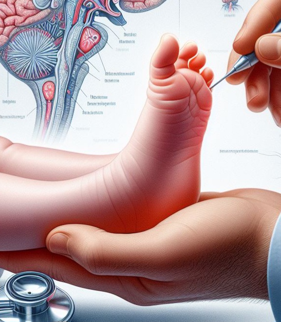
1) Pathologic reflexes of Babinski and Rossolimo groups – in the case of lesions of the corticospinal tract:
|
Reflex |
Procedure |
Response |
|
Babinski |
Stimulate the sole of the foot from heel to toes, in a scratching manner |
Tonic extension of the big toe dorsally, with potential abduction, spreading, and slight plantar flexion of the remaining toes. |
|
Oppenheim |
Apply pressure to the tibia while sliding the thumb and index fingers from a proximal to distal direction. |
-//- |
|
Gordon |
Press the calf muscle area firmly. |
-//- |
|
Schaffer |
Apply pressure to the Achilles tendon using your thumb and index finger. |
-//- |
|
Chaddock |
Gently rub or scratch the area around the outer ankle bone (malleolus). |
-//- |
|
Rossolimo |
Tap or drum the plantar surface of the last phalanges of the toes using your fingers. |
Quick, brief, clonic plantar flexion. |
|
Zhukovsky |
Utilize a neurological hammer to tap the plantar region of the foot, specifically behind the toes. |
-//- |
|
Mendel-Behterev |
Utilize a neurological hammer to tap the os cuboideum on the underside of the sole. |
-//- |
|
Hoffman |
Grasp the middle finger and abruptly pinch its distal phalanx. |
Rapid flexion of all fingers towards the palm (volar flexion). |
|
Tromner |
Grasp the middle finger and tap its distal phalanx from below. |
-//- |
2) Reflexes of oral automatism – in the case of bilateral lesion of the corticonuclear tract
· Labial/snout reflex: Knocking with a neurological hammer on the upper or lower lip causes contraction of m. orbicularis oris, from a mild tremble to complete eminence of the lips.
· Nasolabial reflex: Tapping the posterior aspect of the nose elicits a response similar to the snout reflex.
· Sucking reflex: Touching or gently rubbing the lips with a small brush elicits the same response as the snout reflex.
· Palmomental reflex: Slowly scratching the palm from the wrist to the thumb elicits contraction of m. mentalis.
3) Reflexes of spinal automatism – occur 3 to 4 weeks after a full disruption of the spinal cord above the lumbar intumescence, causing dysfunction in both pyramidal and extrapyramidal systems until the level of the interruption.
· Shortening reflex: Elicited by the physician extending the leg which causes flexion of the hip, knee and ankle joints.
· Lengthening reflex: Elicited by the physician flexing the leg which causes extension of the hip, knee and ankle joints.
· Walking automatism: Stimulation of one foot causes flexion of the same leg and extension of the other leg.
4) Reflexes of brainstem automatism
· Oculocephalic and oculovestibular reflexes:
The oculocephalic reflex is a neurological response observed in humans. It involves the movement of the eyes in response to the movement of the head.
Clinically, the oculocephalic reflex is often tested by rapidly turning the patient’s head from side to side while observing their eye movements.
Results:
- Normal Response: In a normal oculocephalic reflex response, the eyes move in the opposite direction to the head movement. For example, when the head is turned to the right, the eyes should move to the left, and vice versa. Symmetrical and coordinated eye movements in response to head movements suggest intact vestibular function and brainstem integrity.
- Abnormal Response:
- Absent Response: If there is no eye movement in response to head movements, it could indicate dysfunction in the vestibular system or brainstem. This could be due to conditions such as severe brainstem injury, bilateral vestibular dysfunction, or pharmacological suppression of the reflex.
- Asymmetric Response: If there is asymmetry in eye movements, with one eye moving less or in a different direction compared to the other, it could suggest dysfunction or damage to one side of the vestibular system or brainstem. This asymmetry could indicate conditions such as unilateral vestibular dysfunction or brainstem lesions.
- Vertical Eye Movements: In some cases, abnormal vertical eye movements (e.g., nystagmus) may be observed during the oculocephalic reflex test. These abnormal vertical movements could indicate additional vestibular or brainstem abnormalities and may warrant further evaluation.
- Delayed or Disorganized Movements: If eye movements are delayed, jerky, or disorganized in response to head movements, it could suggest dysfunction in the coordination of vestibular and ocular motor systems.
The oculovestibular reflex, also known as the caloric reflex, is a neurological response that involves the coordination between the vestibular system (which helps maintain balance and spatial orientation) and the oculomotor system (which controls eye movements). It is elicited by stimulating the vestibular system through changes in temperature within the inner ear.
Results:
- Normal Response:
- During the irrigation of the ear canal with cold water, the normal response is for the eyes to deviate conjugately towards the side of the stimulated ear. This response indicates intact vestibular function and appropriate coordination between the vestibular and oculomotor systems.
- When warm water is introduced into the ear canal, the eyes should return to the midline position (or opposite side). This response demonstrates inhibition of the vestibular system, which is a normal physiological reaction.
- Abnormal Response:
- Absent Response: If there is no or minimal eye movement in response to caloric stimulation, it suggests dysfunction in the vestibular system or brainstem. This could be due to severe brainstem injury, bilateral vestibular dysfunction, or pharmacological suppression of the reflex.
- Asymmetric Response: Asymmetry in the caloric response, with one eye deviating more or less than the other, indicates dysfunction or damage to one side of the vestibular system or brainstem. This asymmetry could suggest conditions such as unilateral vestibular dysfunction or brainstem lesions.
- Directional Nystagmus: Abnormal eye movements, such as nystagmus (rapid, involuntary eye movements), may be observed during caloric stimulation. The direction and characteristics of nystagmus can provide valuable information about the underlying vestibular pathology.
- Delayed or Disorganized Response: If the caloric response is delayed, diminished, or disorganized, it suggests dysfunction in the vestibular and oculomotor systems.
5) Grasping reflex of Yanishevski
When an object touches the palm of the hand near the metacarpophalangeal area, the patient involuntarily curls all fingers of the hand opposite the cerebral lesion and grasps the object.
6) Central and Peripheral Paralysis Syndromes
Central Paralysis:
- Lesion occurs within the central nervous system (CNS), including the brain or spinal cord – upper motor neurons.
- Etiologies: Strokes, traumatic brain injuries, tumors, neurodegenerative diseases affecting the CNS.
- Manifestations:
- Patterns of weakness often specific to regions controlled by affected CNS areas.
- Often presents with hemiparesis/hemiplegia or paraplegia/quadriplegia.
- Spasticity/rigidity/hypertonia, increased muscle tone, cortical sensation loss.
- Reflexes:
- Usually, hyperreflexia is present.
- Pathological reflexes are present.
Peripheral Paralysis:
- Lesion occurs outside the CNS, involving peripheral nerves, nerve roots, or neuromuscular junctions – lower motor neurons.
- Etiologies: Nerve compression, trauma, infections, autoimmune disorders, metabolic abnormalities.
- Manifestations:
- Weakness or paralysis localized to specific muscles or muscle groups innervated by affected peripheral nerves.
- Distribution follows specific nerve or nerve root territories.
- Decreased muscle tone, flaccidity/hypotonia, peripheral sensation loss.
- Reflexes:
- Hypo- or areflexia is usually present.
- Pathological reflexes are not present.
References:
4. Neurology handbook for medical students. Prof. Penko Shotekov, MD, Ph.D., Sc.D.
Verified by Dr. Petya Stefanova