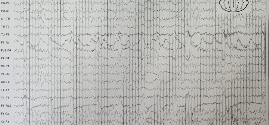
1. What is EEG?
-
- Electroencephalography (EEG) is a non-invasive diagnostic test that evaluates brain wave activity.
- It involves placing multiple small, metal electrodes along the scalp to detect electrical changes in the brain.
- These electrodes pick up signals produced by the brain, which are then recorded by a machine.
2. Purpose and Applications:
-
- Diagnosing Epilepsy: EEG helps diagnose epilepsy by detecting abnormal electrical patterns associated with seizures.
- Other Brain Disorders: It can also identify abnormalities related to other brain disorders.
- Monitoring Consciousness: EEG is useful for evaluating altered consciousness episodes with uncertain causes.
3. Normal EEG Patterns:
-
- The awake EEG typically shows:
- Alpha waves (8-12 Hz, 50 μV) over the occipital and parietal lobes.
- Beta waves (>12 Hz, 10-20 μV) frontally.
- Theta waves (4-7 Hz, 20-100 μV).
- Abnormalities include:
- Asymmetries between hemispheres (suggesting structural disorders).
- Excessive slowing (1-4 Hz, 50-350 μV delta waves) seen in depressed consciousness, encephalopathy, and dementia.
- Abnormal wave patterns, which may be nonspecific or diagnostic (e.g., epileptiform sharp waves).
- The awake EEG typically shows:
4. Activation Techniques:
-
- If routine EEG is normal but a seizure disorder is suspected:
- Hyperventilation, photic stimulation, sleep, and sleep deprivation can help elicit evidence of seizures.
- Nasopharyngeal leads may detect temporal lobe seizure foci.
- Video EEG combines video monitoring with EEG to assess seizure types.
- If routine EEG is normal but a seizure disorder is suspected:
5. Continuous Monitoring:
-
- Ambulatory EEG (over 24 hours or longer) can determine if memory lapses or episodic motor behavior result from seizure activity.
- Video EEG is used before surgery to identify epileptogenic foci.
Remember, EEG is a valuable tool in neurology for understanding brain function and diagnosing various conditions.
References:
1 msdmanuals.com
2 med.uth.edu
3 nhs.uk
4 health.harvard.edu
Verifiziert von Dr. Petya Stefanova
